Gastrectomy

Basic Anatomy of the GI Tract and Accessory Organs

The gastrointestinal system is one long tube from the mouth down. After we ingest food, it is continuously broken down and digested through different chemical and physical processes. The accessory organs of the gastrointestinal system include the tongue, salivary glands, pancreas, liver and gallbladder. Here is a brief outline of the digestive process:


Figure 1: The Gastrointestinal System1

  1. Food enters the mouth, where the teeth and tongue help you chew it into small pieces. Here, saliva partially breaks down some food and helps us swallow it easily.
  2. The bolus (compacted ball of chewed food mixed with saliva) passes through the esophagus, a muscular tube about 25 cm in length.
  3. The esophagus empties into the stomach. Here, strong acids and enzymes chemically break down the food, while stomach muscles act like a blender to break it down even more. This produces a thick slurry known as chyme.
  4. Chyme from the stomach passes into the small intestine where most of the important molecules from the food are absorbed for our body to use. The small intestine is divided into three distinct segments: (1) the duodenum, (2) the jejunum and (3) the ileum.
  5. While in the duodenum, the chyme is exposed to bile (a greenish digestive fluid) secreted by the liver, as well as digestive fluids from the pancreas. These fluids help to further digest the chyme.
  6. The small intestine empties into the large intestine. Here, a lot of the water is reabsorbed and the material is processed by healthy gut bacteria which are important for digestion.
  7. Finally, the waste material is eliminated through the rectum and anus as feces.

If a gastric cancer diagnosis is made early enough, the disease is more likely to be considered curable. This assessment is made based on the stage of the tumor using the TNM system. Surgery for early gastric cancer tries to cure the disease by cutting out the organs affected by cancer cells. There are several different surgical options available to patients and their doctors. Some of the major ones are explained below.

Gastrectomy Part I: The Stomach

Gastrectomy” means to “cut out the stomach”. There are different types of gastrectomy procedures which can be selected based on the location and the extent of a patient’s tumor. Please see Figure 12.

  1. Wedge: the tumor is located in the middle area of the stomach, only along the greater curvature (the left-most edge). Only a triangle-shaped area right around the tumor is cut out.
  2. Central gastrectomy: a band through the middle of the stomach is removed.
  3. Proximal: the tumor is located in the upper stomach only; the part of the stomach closest to the esophagus is cut out.
  4. Distal: the tumor is located in the lower stomach only; the part of the stomach closest to the duodenum is cut out.
  5. Subtotal: the entire stomach except for the very top (which connects with the esophagus and the gastroesophageal junction) is removed
  6. Total: the cancer affects the whole stomach so it must all be cut out. Usually, the lower part of the esophagus and the upper part of the duodenum are also cut out2.  


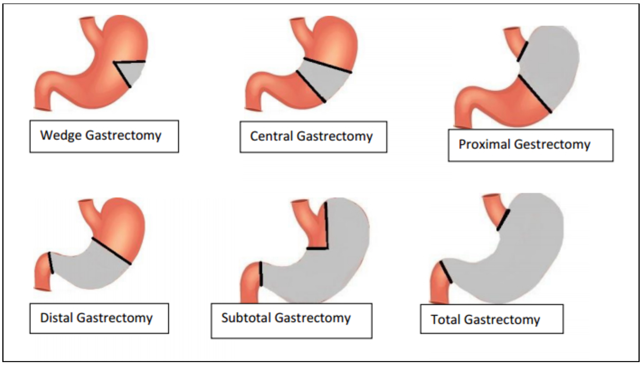
Figure 2: Showing the different regions of the stomach which may be cut out during a gastrectomy surgery; the grey area represents the removed part of the stomach
3

Gastrectomy Part II: Lymph Nodes

Lymphadenectomy” means to “cut out the lymph nodes”. It is also known as a “lymph node dissection”. This is almost always done with a gastrectomy because lymph nodes are a common secondary site for cancer and they provide an easy route for it to spread through the body. In this surgery, lymph nodes are cut out and inspected for the presence of cancer cells.

One of the biggest ongoing debates in gastric cancer treatment amongst experts is what type of lymphadenectomy should be done. See Figure 21

  1. D0: lymph nodes are not removed
  2. D1: this removes the lymph nodes closest to the wall of the stomach only
  3. D1+: this removes more lymph nodes than a D1 resection, but does not touch the nodes along other organs such as the pancreas, as is done in a D2 resection.
  4. D2: this removes the lymph nodes closest to the wall of the stomach and some lymph nodes located further from the stomach near major blood vessels and other organs.
  5. D3: this removes the greatest number of lymph nodes, including those right next to a large blood vessel called the aorta.

It is important to understand that higher numbers correspond to more nodes being removed. For example, a D2 resection removes all the same nodal groups as a D1+ does, plus a few more! Thus, D2 is a more extensive procedure than D1+ is.


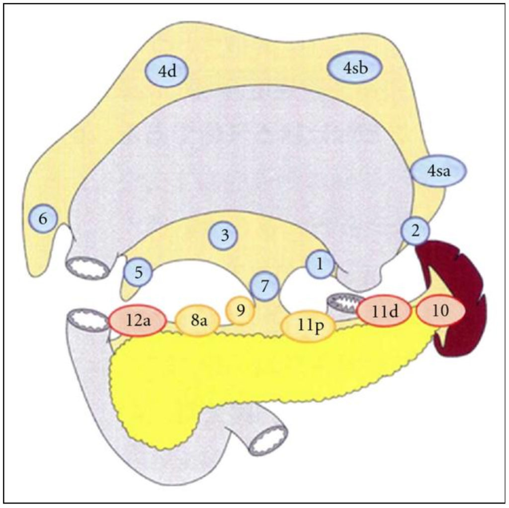
Figure 3: This shows the locations of lymph node groups that are commonly removed in D1 (blue), D1+ (yellow) and D2 (red) lymphadenectomies accompanying a total gastrectomy
2
.

Currently in North America, doctors tend to use the D1 or D1+ technique most often. On the other hand, surgeons in East Asian countries usually use the D2 approach as standard of treatment. Dr. Coburn’s research group has done a lot of work reviewing the lymphadenectomy techniques used in different places, as well as the pros and cons of each approach. Find out more about these studies HERE.

Gastrectomy Part III: Reattachment

Understandably, it may become difficult to eat and maintain a healthy lifestyle after having part (or all) of a major digestive organ removed. If the GI tract is one long tube from the mouth onwards, how can food pass from one side to the other if the stomach is gone? To avoid this problem, the two ends of the GI tract on either side of the cut-out portion must be reattached. There are different techniques that a surgeon can use to achieve this and ensure the continuity of the GI tract. Three of these techniques are explained here3:

  1. Billroth I: also known as a gastroduodenal anastomosis (“anastomosis” means “connection”). If the duodenum is still usable, it is attached to the bottom of the cut portion of the stomach3.
  2. Billroth II: also known as a gastrojejunal anastomosis. Sometimes, the duodenum is not usable because it was also cut out to remove the tumor. In this case, the cut edge of the stomach is attached to the jejunum, which is the second part of the small intestine. It is important to know that the duodenum is the only place where iron is absorbed. So patients whose duodenum is removed are at a greater risk of developing anemia (a condition of low iron in the bloodstream)3.  
  3. Roux-en-Y gastrojejunostomy: this method might be used if a large portion of the stomach and duodenum has been cut out. It attaches the very top part of the stomach (where it connects with the esophagus) to the jejunum3.

All three of these methods work by sealing together the two cut ends of the GI tract, so that food can pass through uninterrupted from the esophagus to the stomach and the small intestine.


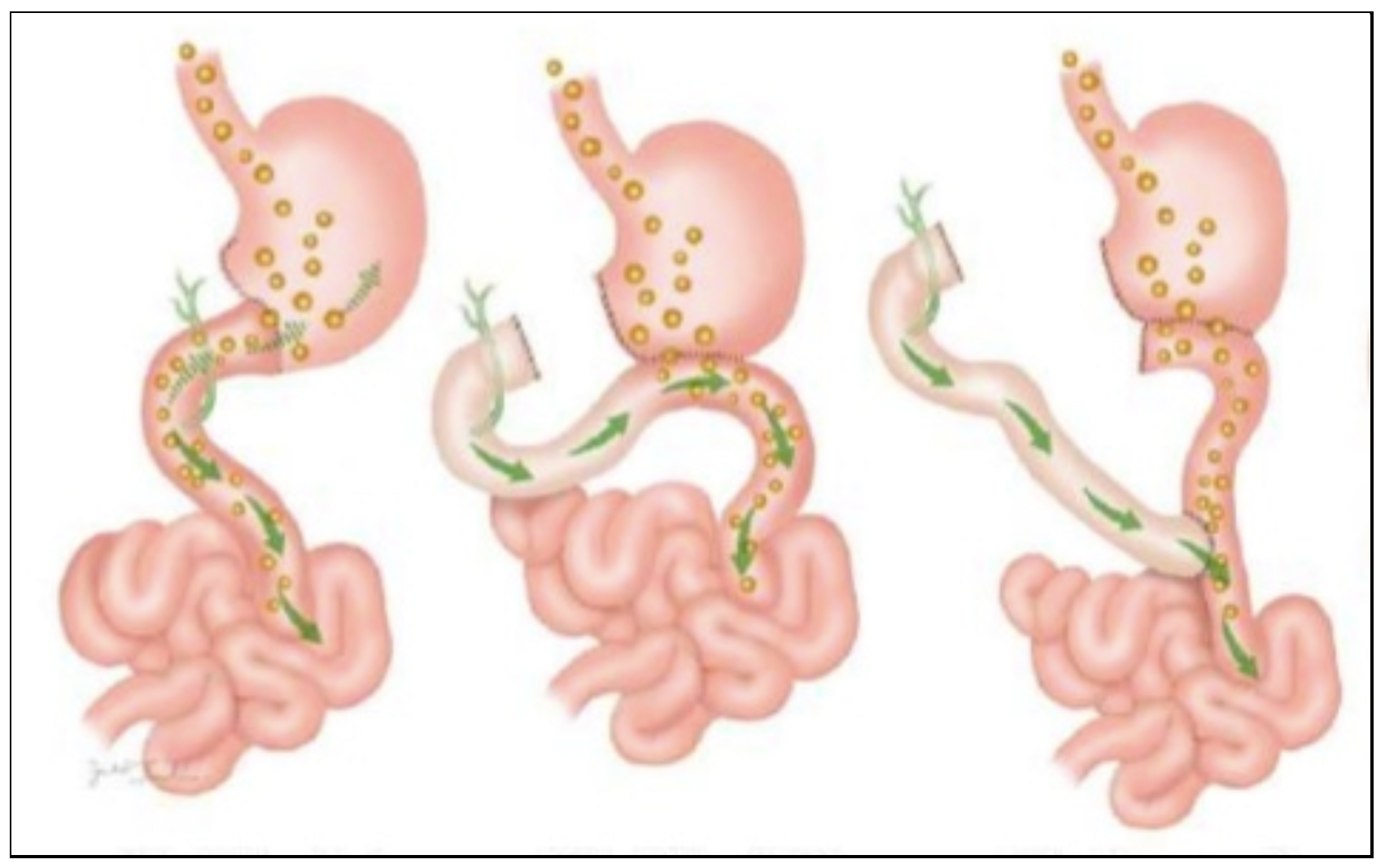
Figure 4: Shows three major reattachment techniques of the stomach to the small intestine after a distal gastrectomy. Far left is Billroth I, middle is Billroth II, far right is Roux-en-Y

Works cited

  1. National Institute of Diabetes and Digestive and Kidney Diseases. Your Digestive System and How it Works. Available from: https://www.niddk.nih.gov/health-information/digestive-diseases/digestive-system-how-it-works . [Accessed: July 2, 2018].
  2. Tamura, S. et al. Lymph node dissection in curative gastrectomy for advanced gastric cancer. International Journal of Surgical Oncology. 2011. Available from: doi: http://dx.doi.org/10.1155/2011/748745.
  3. Virginia Surgery Associates. Gastric Surgery. Available from: https://www.vasurgery.com/surgical-procedures/general-surgery-2/gastric. [Accessed: June 21, 2018].
  4. Zong, L. “Billroth I vs Billroth II vs. Roux-en-Y following distal gastrectomy: a meta-analysis based on 15 studies”. Hepatogastroenterology. 2011. 58:109(1413-1424). Available from: doi: 10.5754/hge10567.

Click here to learn about ‘prehab’ in surgery, and how it helps to prepare your body for gastrectomy.